Tucson Woman Walking Thanks to Thoracic Disc Herniation Surgery
“It was all very sudden,” said Lester, now 45. “I hadn’t had any car accidents, falls, nothing. It was just completely out of the blue.”
In March 2019, she received a devastating phone call about the death of a family member on the East Coast. Suddenly, she felt as if she’d been hit in the back with a rubber band or BB gun.
“I thought it was just stress,” said Lester. She had recently moved from South Carolina to Tucson, Arizona with her teenage son for a new job—only to be laid off six months later.
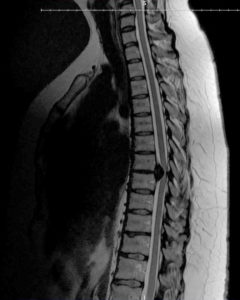
A couple months after the phone call, she started having consistent back pain. It was tolerable, but she grew more concerned when she developed slight numbness in her legs. An MRI revealed a herniated disc in her mid-back, also known as the thoracic region of the spine.
When a Herniated Disc Is Routine—and When It Isn’t
The discs between the bones of the spine (vertebrae) act as shock absorbers and enable the spine to move. Disc herniation occurs when the soft center of a disc pushes through a tear in the tough outer layer and into the spinal canal. The protruding disc fragment can put pressure on the spinal cord, causing back pain and spinal cord dysfunction.
Disc herniation is a common spinal condition that can occur as a result of injury or aging. Over time, intervertebral discs lose their cushioning ability and become more vulnerable to rupture. Research has shown that herniated discs run in families, suggesting a genetic predisposition.
A spine surgeon can remove all or part of the protruding disc in a procedure called a discectomy. If the surgeon removes the entire disc, they will insert a bone or synthetic bone-like material to fuse together the adjacent vertebrae for stability. Metal plates, rods, and screws can help hold the vertebrae together while the bone graft heals.
Calcified giant thoracic disc herniation is one of the most, if not the most, challenging surgical conditions that a spine surgeon can encounter.
-Dr. Juan Uribe, Chief of the Division of Spinal Disorders
Discectomy is a routine spine surgery—unless the disc herniation is in the thoracic region as it was for Lester. The bones that make the thoracic spine more stable and less prone to disc herniation than the lumbar and cervical regions—the ribs and sternum—also make it more difficult for a surgeon to access the region.
A spine surgeon in Tucson discovered through additional tests that Lester’s herniated disc had hardened due to calcium buildup, a process known as calcification, which further complicated her case. When a herniated disc becomes calcified, the spine surgeon must drill the disc until it is paper thin before removing it.

“He said, ‘It’s too big. I’m not going to do this,’” Lester recalled.
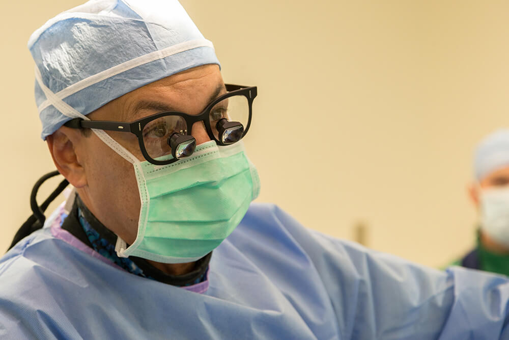
He referred her to Dr. Juan Uribe, chief of the division of spinal disorders and a minimally invasive spine surgeon at Barrow Neurological Institute in Phoenix.
Dr. Uribe told Lester she had the largest calcified thoracic disc herniation he had ever seen.
“Calcified giant thoracic disc herniation is one of the most, if not the most, challenging surgical conditions that a spine surgeon can encounter,” Dr. Uribe said. “The risks associated with the surgical procedure are usually catastrophic, including paralysis, respiratory failure, or vascular injuries.”
But Lester was struggling to walk, as the weakness and numbness in her legs had worsened over the summer.
“Leaving the spinal cord under chronic compression by the herniated disc, or performing partial removal, leads to poor outcomes with significant impact on quality of life,” Dr. Uribe said.
Minimizing Risk with Minimally Invasive Surgery
Traditionally, spine surgeons have accessed the thoracic spine through the front or back of the body, known respectively as the anterior and posterior approaches. The anterior approach requires assistance from a cardiothoracic surgeon and may involve broken ribs and breathing tubes. Posterior thoracic discectomy requires significant muscle cutting in the back, and the delicate spinal cord limits the surgeon’s access to the front of the spine.
Because of the risks associated with the traditional surgeries, Dr. Uribe and other minimally invasive spine surgery specialists developed a lateral (side) approach. They took the extreme lateral interbody fusion (XLIF) procedure used for herniation in the lower back, called the lumbar spine, and adapted it for the thoracic region.
In the lateral thoracic approach, also known as the retropleural approach, the patient lies on his or her side. The spine surgeon makes a small incision over the rib cage and may remove part of the rib. To reach the spinal column, the surgeon gently moves aside the pleura—the tissue that covers the lungs.

Specialized instruments, advanced microscopes, and navigation techniques allow the spine surgeon to work through the small incision. Neurophysiological monitoring records spinal cord function during surgery, reducing the risk of injury to the nerves.
“Being able to access the spinal herniation using less-invasive techniques through a lateral approach, working between the lungs and the dorsal muscles, is advantageous, “Dr. Uribe said. “It potentially prevents chest cavity injuries and posterior hardware placement.”
But even with this less-invasive approach, Dr. Uribe still carefully evaluates each patient and only performs surgery if they are displaying what he calls “red flag signs.” These include difficulty walking, numbness or weakness in the legs, and loss of bowel or bladder function.
Lester was a candidate and decided the possibility of saving her mobility was worth the risks. She leaned on the expertise of Dr. Uribe and on her faith.
“I believe that things happen for a reason,” she said. “How strange is it that I get hired, I move across the country only to lose my job, but yet, I’ve got the nation’s—if not the world’s—best spine surgeon in my backyard?”
An ‘Outstanding’ Outcome
Dr. Uribe performed Lester’s minimally invasive lateral thoracic discectomy in October. Through about a three-inch incision, he removed the disc from her spine. He then inserted a piece of her rib to help fuse the T6 and T7 levels together.
Within 24 hours of surgery, the numbness in Lester’s legs, feet, and toes had almost completely subsided.
“Rebecca’s outcome was outstanding,” Dr. Uribe said. “She was able to leave the hospital and regain her quality of life in a few days.”

Lester returned to work after taking three weeks to recover at home. In addition to her communications job at a cardiology practice, she’s also pursuing her PhD in organizational psychology.
“It’s a testimony to just how skilled and talented Dr. Uribe is,” she said. “I just felt emotionally, psychologically, spiritually, and physically so much better.”
Since her surgery, Lester has shared her story with other Barrow patients who are either recovering from thoracic spine surgery or considering the procedure.
She also says the surgery has given her a new perspective at work. As someone who has always been relatively healthy, she now feels she can relate to patients on a deeper level.
“It’s helped me appreciate everything, just my health and how fortunate I was to be able to move through this so successfully and so quickly,” she said.
