Barrow Study Suggests Immune Cells Contribute to Neurodegeneration
A new study suggests that immune cells in the brain and spinal cord might play a role in neurodegenerative diseases when altered by a specific genetic mutation.
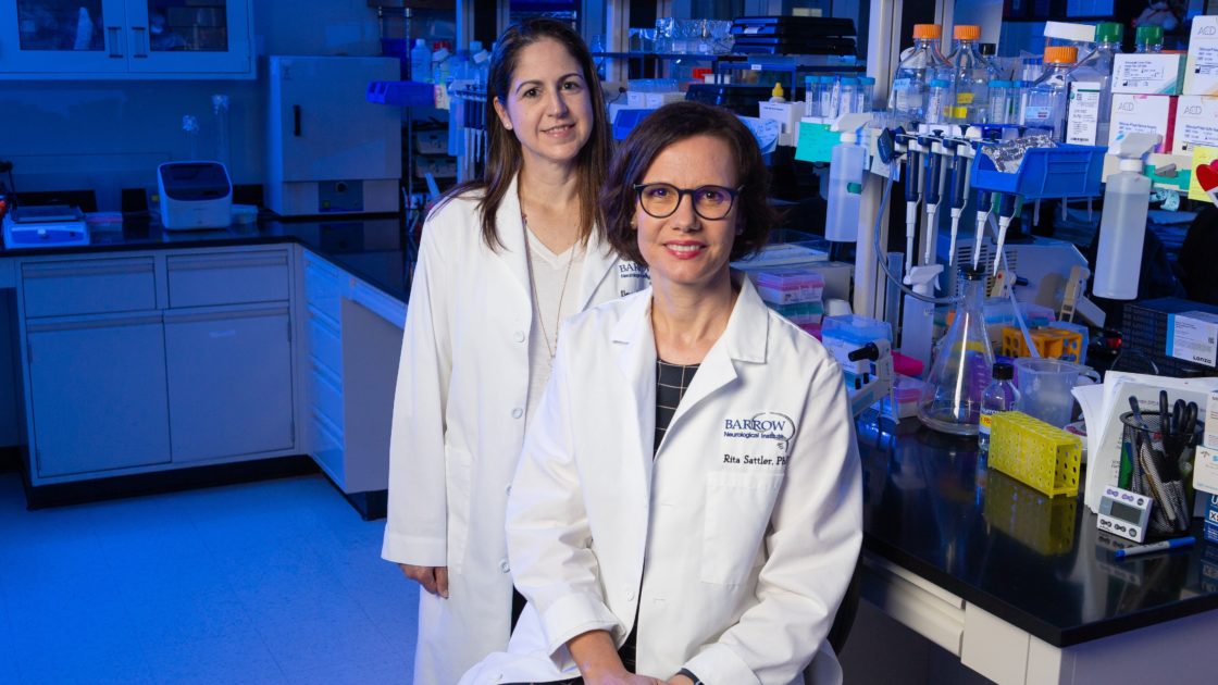
The study, co-led by Rita Sattler, PhD, a professor of neurobiology at Barrow Neurological Institute, was recently published in the journal Neuron.
Mutations in the C9orf72 gene are the most prevalent known cause of amyotrophic lateral sclerosis (ALS) and frontotemporal dementia (FTD). These diseases both involve the deterioration and death of neurons, and they often coexist in patients.
ALS affects motor neurons, which are responsible for controlling voluntary muscle movements. People with ALS gradually lose their ability to move, speak, eat, and breathe. FTD affects the neurons in the brain’s frontal and temporal lobes, causing changes in behavior, personality, and language.
The C9orf72 mutation reduces normal protein function and can instead result in the production of potentially toxic proteins. However, scientists don’t fully understand how this ultimately leads to neurodegeneration—making it difficult to intervene therapeutically.
Microglia in Overdrive
Non-neuronal cells—which do not produce electrical impulses but perform supportive functions—are known to significantly contribute to neuronal loss in neurodegenerative diseases. This most likely occurs early in the disease process.
This study focused on the resident immune cells of the brain and spinal cord, known as microglia. Normally, these cells are the macrophages of the central nervous system. They destroy foreign invaders and clean up accumulating waste left behind by cells performing their normal functions.
During the aging and disease processes, however, microglia appear to become destructive rather than protective.
Investigators observed laboratory mice with normal C9orf72 function and mice lacking the C9orf72 gene, mimicking one consequence of this gene’s disease-causing mutation. The mice depleted of the gene performed significantly worse on a maze test, indicating learning and memory problems.
When the team studied the brains of the two groups of mice, they found that the microglia in those lacking the gene had gone into overdrive.
While clearing waste, these immune cells were also mistakenly eliminating synapses—important cellular structures that enable communication between neurons. Disruptions in neuronal communication can affect normal neurological functions, such as walking, talking, learning, and forming memories.
Ileana Lorenzini, PhD, a postdoctoral fellow in the Sattler Laboratory at Barrow, and her team found that microglia that lacked the C9orf72 gene had an increase in genes associated with a pro-inflammatory, and thereby damaging, immune response.
“More detailed knowledge on how and when the switch occurs in a patient with neurodegenerative disease will facilitate the development of novel therapeutics to treat them,” Dr. Sattler said.
The study also looked at whether the loss of C9orf72 would have an effect on the function of neurons in the presence of amyloid plaques. These are toxic protein accumulations found in the brains of most patients with Alzheimer’s dementia. They are thought to contribute to neurodegeneration.
When researchers genetically removed the gene from mice with amyloid plaques, those mice also had overactive microglia and deficits in learning and memory.
“These studies support a significant role of activated microglia in neurodegenerative diseases and provide new opportunities to therapeutically intervene at an early stage during disease progression, before neuronal loss has occurred,” Dr. Sattler said.
What’s Next?
Dr. Sattler and her laboratory staff are currently expanding on this work in human patient-derived models, which consist of blood or skin cells that have been donated by adult patients and reprogrammed into brain cells.
They are also collaborating with imaging researchers at Barrow to visualize and detect synapse loss in mouse models using positron emission tomography (PET), with the goal of advancing this approach to patients affected by ALS and FTD.
Other staff in the Sattler Laboratory who contributed to this project include Junwon Kim, a freshman at the University of Pennsylvania, and Layla Ghaffari, a neuroscience PhD student at Thomas Jefferson University. This study was done in close collaboration with investigators from Cedars-Sinai Medical Center.


