Clinical Images: Nonsurgical Treatment of a Cervical Epidural Abscess
Paul W. Detwiler, MS, MD
Randall W. Porter, MD
Robert Clark, MD*
Volker K.H. Sonntag, MD
Division of Neurological Surgery, Barrow Neurological Institute, St. Joseph’s Hospital and Medical Center, and *Infectious Disease Consultants, Ltd. Phoenix, Arizona
Key Words: Cervical Epidural Abscess
A 66-year-old retired nurse presented to our Spine Service with worsening cervical pain. A laryngeal carcinoma had been resected 13 years earlier. She subsequently developed esophageal strictures, which were treated with esophageal dilation. The last dilation procedure was performed 6 months before presentation at which time she developed an esophageal perforation and an infection of the cervical prevertebral soft tissue. Biopsy and culture revealed alpha Streptococcus, which was treated with antibiotics for 4 months.
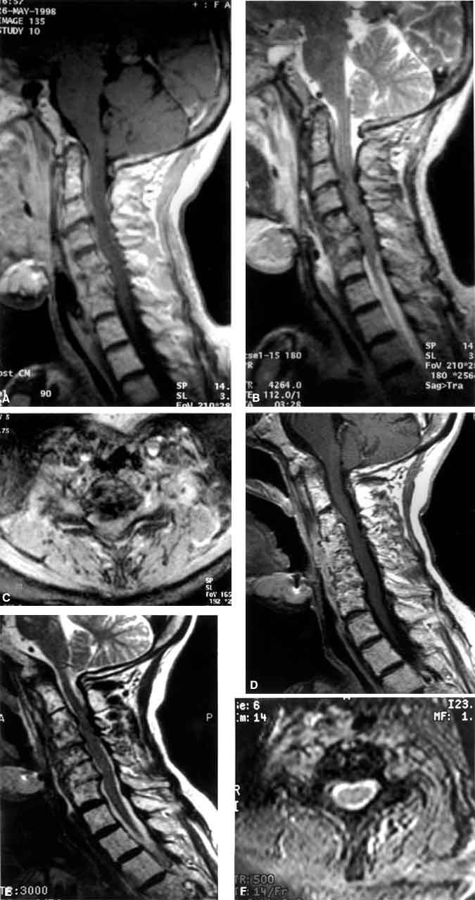
She presented to our service with worsening cervical pain, 4-/5 weakness throughout the extremities, and stocking-glove numbness and tingling in the hands and feet. A sagittal T1-weighted magnetic resonance (MR) image of the cervical spine with contrast (Panel A) demonstrated destruction of the C5-C6 and C6-C7 disc spaces and adjacent vertebral bodies. Swelling of the prevertebral soft tissue and significant enhancement of the ventral epidural space consistent with an abscess were visible. A sagittal T2-weighted MR image (Panel B) showed a loss of signal intensity from the cerebrospinal fluid (CSF) around the spinal cord from C3 to C6. An axial T2-weighted MR image at C5-C6 (Panel C) demonstrated complete loss of signal intensity from the CSF with ventral compression of the spinal cord.
The patient was offered conservative treatment with intravenous antibiotics or anterior decompression and debridement, fusion, and plating. Because of severe anterior and posterior cervical scarring, we were concerned that her risks for postoperative wound dehiscence or infection were high. She selected the conservative course. She was immobilized in a halo brace and treated with metronidazole, clindimyacin, vancomycin, and ceftazidime.
She underwent follow-up magnetic resonance (MR) imaging after a 5-month course of intravenous antibiotics. A sagittal T1-weighted MR image with contrast (Panel D), a sagittal T2-weighted MR image (Panel E), and an axial T2-weighted MR image (Panel F) at C5-C6 showed that the swelling of the prevertebral soft tissue had resolved. There was destruction of the intervertebral disk spaces at C3-C4, C4-C5, C5-C6, and C6-C7. The ventral epidural enhancement had disappeared. The axial T2-weighted image (Panel F) demonstrated CSF around the entire spinal cord. The patient’s strength improved to 5/5 in all extremities. She will be followed closely for recrudescence of the osteomyelitis as well as for delayed cervical kyphosis.

