
CT Angiography and Stroke
Thomas A. Yeo, MD
Robert Wallace, MD
Shahram Partovi, MD
Division of Neuroradiology, Barrow Neurological Institute, St. Joseph’s Hospital and Medical Center, Phoenix, Arizona
Abstract
In the clinical setting of acute stroke symptoms, rapid and accurate diagnostic imaging is critical for treatment evaluation. In addition to routine brain computed tomography (CT), CT angiography of the cervical and cerebral circulation can rapidly contribute valuable additional diagnostic information. Not only can the site of occlusions in major branch vessels be identified, but the status of collateral blood flow and injured but potentially salvageable brain parenchyma can be assessed. A valuable roadmap of the arterial vasculature is obtained, and decisions about the need for endovascular treatment can be made quickly. Even if urgent interventional care is unnecessary, CT angiography answers questions about the possible etiology of a patient’s symptoms and guides future medical and surgical treatment decisions.
Key Words: carotid stenosis, CT angiography, intracranial stenosis, stroke, thrombolysis, vertebral stenosis
Stroke is a major cause of death and complications—the third leading cause of death behind coronary artery disease and cancer. The theme common to treatments for acute stroke is rapid revascularization to restore blood flow to the ischemic brain quickly enough to minimize the size of the stroke. This goal requires obtaining a fast and accurate diagnosis from clinical and imaging information. The patient’s medical history and neurologic examination are the key elements in the initial diagnosis of stroke.[15] Appropriate clinical inclusion and exclusion criteria must be used to reduce the risks of reperfusion injury, most importantly, hemorrhage.[10] Clinical information about the time of stroke onset, severity of neurological impairment, complicating medical conditions, or factors that predispose to hemorrhage is necessary to move to the next phase of the diagnostic evaluation.
If clinical criteria demonstrate that the stroke patient may be a candidate for acute intervention, obtaining fast and accurate imaging information is the next critical step.[5] The most important imaging questions that need to be answered to guide rapid treatment are as follows: Is the lesion hemorrhagic? Is there another explanation for the patient’s condition such as a mass? Is there already evidence of significant brain injury resulting from an ischemic event that would preclude safe revascularization?
Few would argue that magnetic resonance (MR) imaging, including perfusion and diffusion imaging, is superior to computed tomography (CT) in detecting subtle and early infarcts. CT, however, is more widely available, faster, typically less expensive than MR imaging, and extremely sensitive in detecting acute hemorrhage. CT scanners often are located closer to emergency departments than MR imaging units, allowing easier access to support personnel and equipment. Furthermore, all major clinical stroke trials to date have used CT before patients were randomized to treatment groups. Consequently, CT is the most common first choice for imaging stroke patients. By combining CT angiography with routine brain CT, as discussed in this article, important information about the cerebral circulation for the assessment of acute stroke patients can be obtained rapidly.
Angiographic Options in Stroke Imaging
Although CT angiography shares a common suffix with MR angiography and digital subtraction angiography (DSA), the differences among these techniques are far more compelling than this superficial similarity. There are at least two major differences among these angiographic techniques. First, the type of information gathered differs. Second, how this information is presented to the observer (via postprocessing) is unique to each modality.
Digital Subtraction Angiography
DSA is the angiographic gold standard against which other angiographic modalities are judged in terms of accuracy and image representation. During a DSA study, intra-arterial x-ray contrast media is injected intravenously at a high rate, and images of the area of interest are acquired in rapid succession. Before the bolus of contrast reaches the brain, several noncontrast images are acquired first. These initial images serve as “masks” that are used to subtract soft tissue and osseous structures from the final angiographic images. After the subtraction process, the only identifiable structures are vascular and contain the radiographic contrast material within their lumen.
Numerous images are acquired in sequence, in effect, creating a movie that displays the passage of the contrast bolus through the arterial tree, capillary bed, and, finally, into the venous phase. In this manner, DSA measures not only the physical inner luminal diameter of a vessel but also provides information about the rate of blood flow through the vascular structures. DSA does not measure the outer diameter of a vessel directly. However, indirect evidence, such as displacement of vessels via mass effect, may be present. Hence, a vascular abnormality seen on DSA may represent only a small visualized portion of a much larger unopacified vascular abnormality.
Moreover, DSA is an invasive method for assessing the vasculature of the brain. It requires an arterial puncture with a large bore needle and the introduction of a flexible catheter into the carotid arteries. In the United State, the risk of stroke during this procedure is 0.3% to 1.2%.[3,6,12] Catheter-related vascular dissections and allergic reactions to the contrast media pose additional risks.
Finally, it is noteworthy that DSA is a two-dimensional technique. At a minimum orthogonal views are necessary, and multiple views are needed to profile a given lesion or vascular territory. Currently, rotational three-dimensional (3-D) techniques are only available with prolonged, slow injections of contrast media. These techniques are used to focus an evaluation on problematic vascular anatomy. They do not provide the series of images through the entire circulation needed to derive significant physiological information about circulation time.
MR Angiography
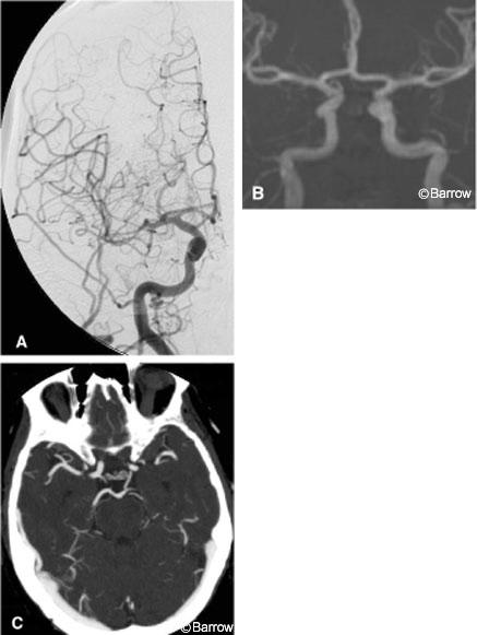
MR angiography is a noninvasive cross-sectional angiographic method that has gained widespread acceptance for the diagnostic imaging of stroke because of its high sensitivity and specificity and its ability to emulate DSA visually. This visual similarity has helped create a relatively seamless transition from DSA to MR angiography for both radiologists and clinicians (Fig. 1).
Despite their apparent visual similarity, MR angiography and DSA measure very different parameters. Unlike DSA, MR angiography is not a direct measure of the actual luminal diameter. Rather, it is an angiographic image derived from the movement of water protons through a vessel. MR angiography is exquisitely sensitive to the rate of proton movement. If the rate is too fast or too slow (i.e., jet flow or sluggish flow), the signal from the moving proton can lead to a false reading as happens in a flow gap. False readings are not necessarily problematic in MR angiography for they can indicate an underlying disease. For example, a flow gap can signal the presence of severe underlying vascular stenosis with associated jet flow. In contrast, a vessel has an identifiable physical lumen on DSA regardless of the rate of blood flow.
Relative and absolute contraindications primarily relate to issues of the magnetic field in use. Perhaps the greatest limitation is the need for patients to remain motionless for 10 to 15 minutes depending on the protocol used. Artifact from patient movement can markedly decrease the sensitivity and specificity of an MR angiographic examination. While DSA can identify the direction of blood flow, MR angiography requires some a priori suspicion that the direction of blood flow is abnormal to enable detection because special techniques and parameters must be used.
CT Angiography
The main advantage of CT angiography is that it can be performed quickly and reliably at the time of a patient’s initial CT study. The success of MR angiography in visually simulating DSA has translated into an unfortunate disadvantage for CT angiography. As the newest cross-sectional member of this diagnostic family, it too is expected to provide images that are visually similar to DSA.
Indeed, some have tried to make CT angiographic images look like MR angiography and DSA. However, most investigators agree that multiplanar volume reconstruction (MPVR) images are the most efficacious mode of viewing CT angiograms. Consequently, one challenge is to persuade users to accept this new method of display.
Despite its apparent visual dissimilarity with DSA, CT angiography is functionally closer to DSA than MR angiography. CT angiography measures the finite physical lumen of a vascular structure and thus is more like DSA than MR angiography. Essentially a snapshot of contrast-filled vascular structures at one point in time, CT angiography provides no direct information about rate or direction of blood flow. It is possible to hypothesize about rate and direction of blood flow based on a general understanding of human physiology. These assumptions, however, would have to be verified if treatment would be altered as a result. On the other hand, the physiological aspects of turbulent blood flow will not cause the morphology of a vessel to be miscalculated on CT angiography as it can be on MR angiography. If the administration of a bolus of contrast is timed appropriately, the filled structures are mostly arteries but mixed arterial and venous imaging is common. CT angiography exceeds the capabilities of DSA in that it provides direct information about the outer luminal diameter of a vessel.
During CT angiography, contrast media is injected into an antecubital vein. Therefore, the need for an arterial puncture and the risks of catheter-related strokes and dissections are eliminated. However, the risk of contrast-related reactions is higher with intravenous injections than with intra-arterial injections. A high rate of flow (4 ml/sec) is required to keep the contrast bolus “tight.” Thus, the intravenous site must be scrutinized to avoid subcutaneous extravasation of the contrast media. Scanning the brain is limited to the circle of Willis and begins once the bolus reaches the skull base. The wave of bolus is followed as the contrast media passes rostrally through the brain during the arterial phase.
Timing the scan to match the passage of the contrast bolus is critical. Various techniques have been proposed to help time the administration of the contrast, but the ideal solution has yet to be developed. Most institutions use a fixed timing delay to reduce operator-dependent and other potential errors. A delay of 9 to 16 seconds with an 80 to 100 ml bolus of contrast typically provides excellent arterial opacification with minimal venous “contamination.” If a patient’s cardiac output is greater than normal, venous opacification can be expected to increase. If a patient’s cardiac output is unusually low, the scan may begin before the bolus arrives and arterial opacification will be insufficient. A “missed bolus” means that a patient has been exposed fruitlessly to radiation and contrast media and their associated risks. These pitfalls related to timing represent a final obstacle to the widespread use of CT angiography.
The width of CT angiographic images ranges from 0.6 to 2.5 mm. Multidetector technology allows four or more images to be acquired per CT rotation. This ability to acquire multiple images is critical to CT angiography where speed is essential. With CT angiography rapid acquisition is not simply a luxury; it is a necessity to avoid venous contamination and the superimposition of venous structures.11 With a total circulation time of about 6 seconds in the brain, the scanner has only 3 seconds to image from the skull base to the pericallosal vessels during the arterial phase. Consequently, the slower that the rate of scanning is, the greater will be the extent of venous contamination.
Occasionally, venous opacification obscures adjacent contiguous arterial structures. In our experience, this problem can be particularly challenging during the evaluation of the cavernous segment of the ICA because of opacification of the surrounding cavernous sinus and sylvian portions of the middle cerebral arteries (MCAs), which are close to numerous venous structures in the sylvian fissure. Atherosclerotic calcification, which is common in the carotid siphon, further compounds the difficulty of evaluating this region. Delayed circulation times or too short of a programmed scan delay can have the opposite effect: The area of interest may be scanned before the bolus of contrast has opacified the target vasculature completely.
Newer generation CT scanners acquire eight slices per rotation, and each rotation lasts 0.5 to 1.0 seconds. Ultimately, 16 scans per rotation and more are expected to be achieved. With each iteration of scanners, CT angiography will decrease the amount of associated venous contamination. With sufficiently fast multidetector scanners, CT angiography theoretically could be followed immediately by CT venography by scanning the same bolus as it passes beyond the arteries into the venous phase. The possibility of dural sinus thrombosis could then be assessed.
Scan artifact from adjacent bone or vascular clips can be troublesome. Surgical clips and other metallic objects can cause significant beam-hardening artifacts that obscure adjacent vessels. Occasionally, widening the settings of the viewing window can improve visualization of the vessels, leading to a more confident assessment of the vasculature. For the same metallic object, the degree of artifact associated with CT angiography is often significantly less than that associated with MR angiography (see CT Angiography: A Tool for Management of Cerebral Aneurysms? Fig. 8).
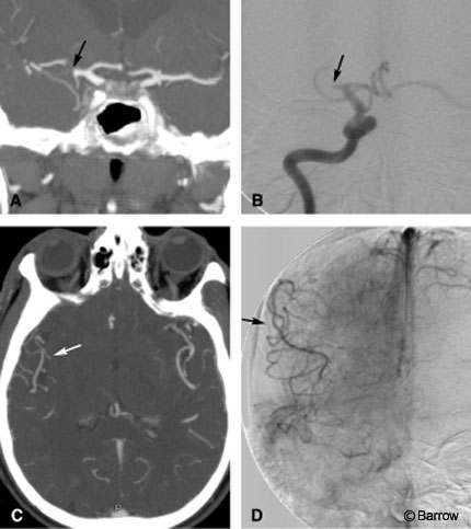
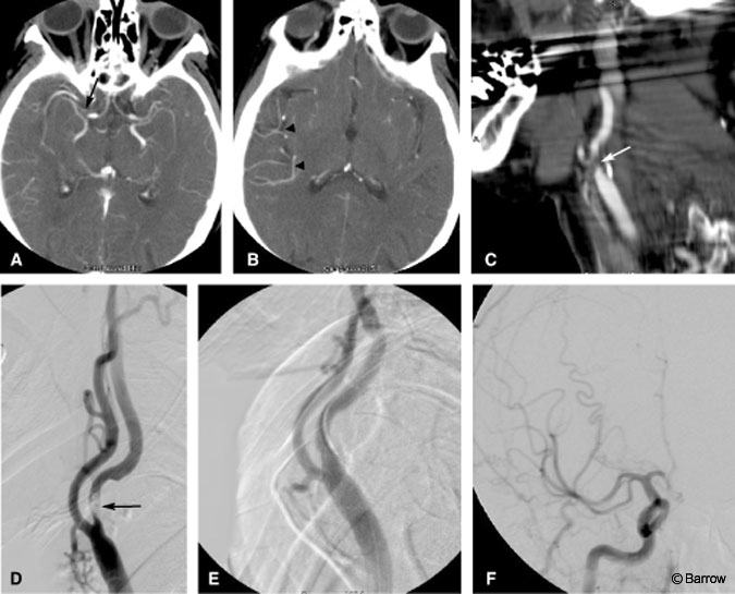
in the right hemisphere despite the occluded right M1. We hypothesized that these vessels were filling in a retrograde fashion via leptomeningeal collateral circulation. (D) Late arterial DSA confirmed retrograde filling (arrow) of the right hemispheric branches via collateral pathways.
CT Angiography of the Brain
In the setting of acute stroke, CT angiography provides a map of the vasculature that can influence treatment strategies.[14] CT angiography can diagnose occlusion of all large intracranial arteries with high rates of both sensitivity and specificity.[13] Shrier et al.[11] reported an overall agreement of 99% between CT angiography and DSA used to evaluate intracranial stenosis or occlusion. Na et al.[9] identified acute proximal occlusion of the MCA using CT angiography in 17 of 18 patients, and Knauth and coworkers7 identified 100% of patients with large proximal occlusions of the ICA, MCA, and basilar trunk (Fig. 2). As the site of occlusion becomes more distal, it becomes more difficult to demonstrate reliably the site of branch vessel thrombus. To help gauge potential candidates for thrombolysis, the volume of tissue compromised by hypoperfusion can be estimated.
Acute MCA occlusion is the most common cause of cerebral infarction. The Prolyse in Acute Cerebral Thromboembolism (PROACT) study was a multicenter randomized controlled trial using cerebral angiography and intra-arterial thrombolysis to treat acute MCA occlusions.[2] The PROACT trial is the only multicenter randomized trial, in which cerebral angiography was performed at the time of acute stroke treatment, and the findings supported clinical suspicions. In the PROACT study, 38% of patients who underwent angiography had an occlusion of the first (M1) or second (M2) segment of the MCA. Twenty percent presented with ICA occlusion instead of MCA occlusion, 19% had no occlusion at all, and 8% had distal MCA branch occlusions that were unamenable to transcatheter treatment. These patients were evaluated by conventional angiography. The findings suggested that a negative CT angiographic screening evaluation could eliminate the need for conventional angiography and prevent the expensive deployment of the endovascular team.
The initial vessel targeted for evaluation through DSA is based on the stroke patient’s presenting symptoms. The necessity for initiating urgent therapy usually precludes a complete prethrombolysis catheter angiographic evaluation of the collateral circulation. CT angiography, however, can provide important information about the rest of the cerebral circulation before endovascular treatment begins. For example, CT angiography adequately visualizes leptomeningeal collateral blood flow, as demonstrated by enhancing MCA branches beyond the site of arterial occlusion (Fig. 2). Knauth et al.[7] reported an interobserver agreement of 88% when CT angiography was used to evaluate the leptomeningeal collateral blood supply in 17 occlusions of the anterior cerebral circulation.
Predicting the volume of infarcted tissue can be difficult when good collaterals, as defined by filling of the MCA branches in the sylvian fissure, are visualized. The hypodense parenchymal changes seen on CT, combined with the site of vessel occlusion, leptomeningeal collateral flow, and nonenhancing parenchyma, all help predict the size of infarcted and ischemic but potentially salvageable tissue.[8] When CT perfusion imaging is added to the CT angiographic protocol, the volume of ischemic tissue can be estimated even more accurately (see CT Perfusion Imaging, Illustrative Case 1). Recanalization of occluded arteries is more likely to be beneficial when the size of the hypodense and nonenhancing parenchyma is smaller than the territory of the occluded vessel.[7]
CT Angiography of the Neck
The complete vascular evaluation of patients with an acute cerebral infarction typically includes the cervical carotid and vertebral arteries. Cervical carotid stenoses, thrombi, occlusions, and dissections can lead to acute infarctions, and each can be identified by CT angiography in the acute setting. Our CT angiographic protocol for acute stroke often includes evaluation of the cervical vasculature from the aortic arch rostrally to the skull base.
Cervical Carotid Stenosis
The correspondence between CT angiography and DSA for detecting and grading carotid stenosis has been reported at 94%.[1] CT angiography demonstrates the greatest degree of stenosis on axial images. Therefore, axial source images should be reviewed closely. CT angiography allows the user to calculate the cross-sectional area of a stenotic segment. If desired, the ratio of areas between stenotic and normal segments can be calculated, simulating the North American Symptomatic Carotid Endarterectomy Trial method used to estimate stenosis on DSA. Typically, DSA obtains information from only two to three planes, and the stenosis is graded based on the view that indicates the greatest degree of narrowing. Consequently, conventional DSA can underestimate the severity of stenosis if the worst possible view was by chance not obtained.
Rotational and 3D angiography often depict more severe ICA stenosis than conventional biplanar DSA. In this method numerous projections are obtained as the x-ray tube rotates about the area of interest, thereby improving identification of the projection with the greatest narrowing.[4] Because the MR angiographic signal is related to blood flow and is not a direct measure of luminal diameter, stenosis can be overestimated in instances of rapid turbulent flow. Newer techniques and better MR equipment have helped to minimize such flow-related artifacts.
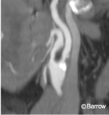
Calcifications of the carotid bifurcations are better visualized on CT angiography than on DSA or MR angiography (Fig. 3).[1] As in all CT examinations, images must be viewed at appropriate settings for window width and level to differentiate contrast opacification from calcifications. In most cases, evaluating carotid narrowing in all three planes at optimal settings for window width and level will depict the degree of stenosis despite dense adjacent calcifications.
If flow is too slow, as in severe carotid stenosis (i.e., the “string sign”), MR angiography can fail to demonstrate the presence of flow not only at but also beyond the level of stenosis. The distinction between a string sign and complete occlusion in the acute setting is important because the former may be treated by an acute thromboendarterectomy while the latter is managed medically.
Illustrative Case 1
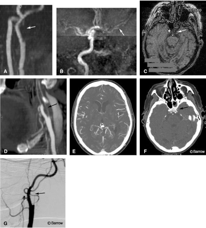
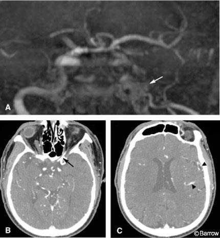
A 63-year-old man presented to the emergency department with slurred speech. His initial CT examination was normal as was a basic MR imaging study of the brain. MR angiography of the neck, however, revealed the absence of a flow signal in the left ICA (Fig. 4A) while MR angiography of the brain showed significantly attenuated signals from left hemispheric branches of the MCA and anterior cerebral artery (Fig. 4B). Axial source MR angiograms also failed to show any detectable flow signal (Fig. 4C). Treatment hinged on excluding a string sign. CT angiography of the neck clearly showed the presence of blood flow in the left ICA, and an MPVR image clearly showed a string sign (Fig. 4D). CT angiography of the brain revealed a significant amount of blood flow in the left hemisphere (Fig. 4E). Axial source CT angiograms confirmed the presence of contrast in a string-like ICA (Fig. 4F). Before surgical intervention, dynamic assessment of a possible external carotid artery-to-ICA anastomosis was performed, and this DSA study again showed the string sign (Fig. 4G).
This case demonstrates the significant role CT angiography can play in differentiating complete ICA occlusion from a string sign. Because CT angiography identifies the presence of a physical lumen in a vessel, it can distinguish a string sign from complete occlusion more reliably than MR angiography. Not only is this distinction important for treatment planning, it also obviates the need for an urgent diagnostic DSA. The small but tangible risk of further procedural complications is thereby avoided. In a number of similar cases at our institution, CT angiography showed the presence of slow blood flow while MR angiography suggested complete occlusion. Furthermore, the difference in scan time between the two modalities was dramatic. Motion artifact during MR angiography of the brain was significant. The MR angiographic scan required about 12 minutes, and the patient was unable to remain motionless for the duration of the study. In contrast, CT angiography of the brain was acquired in less than 60 seconds during which the patient did not move.
False-Negative String Sign on CT Angiography
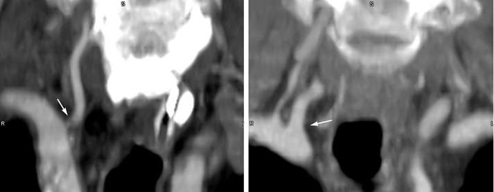
Conceivably, a carotid stenosis could be so severe that the flow of contrast is extremely slowed. An unusually long transit delay for the bolus could then lead to a missed diagnosis if CT angiographic scanning begins before the contrast passes into the poststenotic segments (Fig. 5). This pitfall can be overcome by obtaining a delayed scan through the neck if conventional CT angiography suggests complete arterial occlusion. If the delayed images show the presence of contrast above the suspected level of occlusion, the diagnosis would be altered from complete occlusion to preocclusive stenosis.
In the event of complete carotid occlusion, CT angiography also can help assess the collateral circulation (Fig. 6). Although collateral reconstitution of carotid flow is better depicted by the dynamic imaging of DSA, these collateral pathways are visible on CT angiography if the images are evaluated carefully. It is difficult to evaluate small collateral vessels on MR angiography if blood flow is slow. Our CT angiographic protocol for suspected preocclusive stenosis or string sign includes delayed scanning through the neck immediately after conventional CT angiography of the neck. If intravascular contrast is seen on the delayed scan but not on the initial scan, preocclusive stenosis is suspected.
Tandem Lesions
CT angiography also can detect large ulcerations and tandem lesions. A filling defect within an opacified vessel lumen suggests soft plaque or possible thrombus. Sagittal and coronal MPVR images are especially useful in identifying potential ulcerations. Typically, a second more distal ICA stenosis, known as tandem stenosis, is easily identified on DSA but can be overlooked completely on MR angiography if blood flow is reduced significantly by disease at the bifurcation. The presence of tandem lesions is important because the reduction in blood flow caused by two significant lesions is greater than would result from either lesion alone. Their presence can affect surgical decision-making.
When CT angiography of the brain leads to intra-arterial thrombolysis, CT angiography of the neck helps the interventionist to determine the best vascular route to the offending lesion. Occasionally, CT angiography reveals proximal stenosis, thrombus, or an unusual vascular loop that can alter the planned endovascular pathway. Traditionally, this information would have been available only after an arterial puncture had been performed and preliminary angiographic images had been acquired.
Illustrative Case 2
A 71-year-old woman presented to our emergency department with an acute left hemiparesis of 2 hours duration. Initial noncontrast CT of the head was normal. CT angiography of the neck and brain was performed according to our stroke center protocol. The former identified a clot in the right MCA just distal to the origin of the right M1 (Fig. 7A). The presence of contrast distal to the clot in the right MCA was assumed to represent collateral blood flow in a retrograde fashion. On more cephalad images, the right hemispheric branches were more conspicuous than the left, suggesting slow or delayed retrograde flow (Fig. 7B). CT angiography of the neck identified an intravascular filling defect in the right proximal ICA compatible with an acute thrombus (Fig. 7C). Intravenous tissue plasminogen activator was begun, and the patient was rushed to the interventional suite. The proximal lesion (Fig. 7D) was stented acutely (Fig. 7E) to gain rapid access to the intracranial vasculature for intracranial thrombolysis (Fig. 7F).
This case demonstrates the potency of CT angiography for diagnosing proximal vascular occlusion and for helping to plan therapeutic intervention. The identification of a large proximal lesion alerted the interventionalist to the possible necessity of stenting before thrombolysis. This a priori information helped to expedite the procedure by allowing early preparation and selection of equipment.
Vertebral Artery Stenosis
CT angiography can contribute to the noninvasive assessment of concurrent lesions at the origin of the great vessels of the neck. Traditionally, DSA has been used to identify stenosis of the origin of the vertebral artery. Now, however, stenosis of the vertebral artery, particularly involving its origin, often can be evaluated with less invasive CT angiography (Fig. 8). MR angiography of the origins of the vertebral artery is degraded by respiratory motion because of the time needed to gather the data. Compared to noncontrast MR angiographic sequences, gadolinium contrast-enhanced MR angiography may improve the accuracy of imaging stenosis of the vertebral artery. CT angiography renders respiratory motion less of a problem than with MR angiography because of the speed of data acquisition.
As in the carotid artery, if images are acquired before the contrast arrives, it becomes impossible to interpret vascular disease. This situation is most frequently encountered when evaluating diseases of the origin of the vertebral artery because scanning low in the neck occasionally begins before the arterial bolus arrives. This likelihood is less problematic higher in the neck because the contrast typically catches up with the scanning at the level of the carotid bifurcations.
Challenges to the imaging and interpretation of disease involving the origins of the vertebral arteries on CT angiography include beam-hardening artifact from the shoulders, clavicle, dental work, and metallic clips or metallic foreign bodies; a large body habitus; and even residual contrast in the subclavian vein. Contrast can even flow retrograde in the valveless venous structures of the neck and potentially overwhelm the lumen of the small adjacent proximal vertebral artery. Nonetheless, our experience suggests that these artifacts are less limiting than those encountered in the same area with noncontrast MR angiography. Artifacts from dense vascular calcifications also occasionally limit evaluation of the degree of carotid or vertebral stenosis.
Finally, the origins of other great vessels are often easier to visualize than that of the vertebral arteries because the diameter of the former is larger. The pitfalls are similar. If the origins of the vertebral arteries are visualized on a study, the origins of other vessels also will likely be visible.
Arterial Dissections
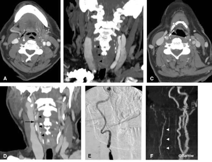
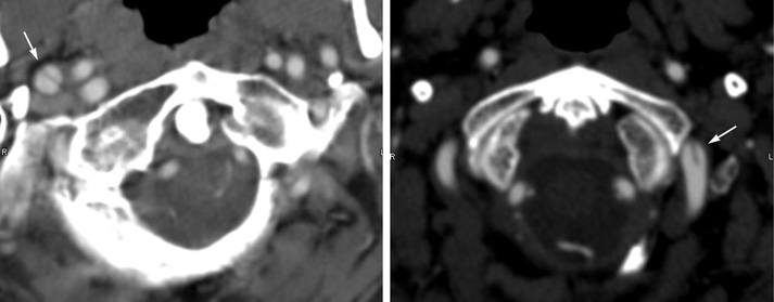
In younger populations, the etiology of stroke includes a greater percentage of extracranial carotid and vertebral artery dissections than found in older populations. Pathologically, blood penetrates the arterial wall, narrowing or occluding the arterial lumen and sometimes enlarging the external diameter of the vessel. Common causes of dissections include trauma, underlying vasculopathy (e.g., fibromuscular dysplasia), hypertension, certain drugs, and vigorous physical activity. CT angiography can diagnose arterial dissections in the cervical circulation just as it can in the aorta. Intimal flaps are visible when contrast-enhanced blood within true and false lumens opacifies both sides of the dissected intima (Fig. 9). Subintimal hematomas appear as filling defects that smoothly narrow the arterial lumen. Although these findings are likely better appreciated on DSA, CT angiography can show the enlarged external diameter of the vessel, thus identifying the occasional dissection that does not narrow the lumen. Fat-suppressed T1-weighted MR images also are useful for detecting dissections, especially when blood is in the subacute stage (i.e., methemoglobin) and appears hyperintense on T1-weighted images. Acute hemorrhage (within the first two days) potentially could be missed on MR imaging because the dissected blood is isointense to the surrounding tissue.
Conclusion
In the event of suspected acute cerebral infarction, CT is the most widely used initial diagnostic tool. Historically, the only protocol offered by CT scanners for assessing such patients was a conventional noncontrast CT examination of the brain. If the head and neck vasculature had to be evaluated, all options involved transporting the patient to another diagnostic room and valuable time for salvaging dying brain tissue was lost. Advances in CT equipment have now increased options for patients in the CT suite. The need to relocate patients and lost time are avoided. These features enhance both speed of diagnosis and the overall quality of care.
CT angiography is a safe, rapid, and accurate diagnostic tool for evaluating acute stroke patients, especially combined with routine brain CT and CT perfusion imaging. CT angiography not only identifies the site of vascular occlusion, it also can determine the degree of leptomeningeal collateralization and help to estimate the amount of hypoperfused brain parenchyma. When this information is combined with information from the cervical circulation, the appropriate treatment can be selected and rapidly instituted. If endovascular treatment is contemplated, CT angiography can be used as a roadmap to plan the procedure. Alternatively, when CT angiography is negative, it may prevent unnecessary urgent conventional angiography.
References
- Bozzao A, Floris R, Villani A, et al: An evaluation of the carotid bifurcation and of the intracranial circle by angio-spiral computed tomography [Italian]. Radiol Med (Torino) 95:577-582, 1998
- del Zoppo GJ, Higashida RT, Furlan AJ, et al: PROACT: A phase II randomized trial of recombinant pro-urokinase by direct arterial delivery in acute middle cerebral artery stroke. Stroke 29:4-11, 1998
- Dion JE, Gates PC, Fox AJ, et al: Clinical events following neuroangiography: A prospective study. Stroke 6:997-1004, 1987
- Elgersma OE, Buijs PC, Wust AF, et al: Maximum internal carotid arterial stenosis: Assessment with rotational angiography versus conventional intraarterial digital subtraction angiography. Radiology 213:777-783, 1999
- Hacke W, Kaste M, Fieschi C, et al: Intravenous thrombolysis with recombinant tissue plasminogen activator for acute hemispheric stroke. The European Cooperative Acute Stroke Study (ECASS). JAMA 274:1017-1025, 1995
- Heiserman JE, Dean BL, Hodak JA, et al: Neurologic complications of cerebral angiography. AJNR Am J Neuroradiol 15:1401-1411, 1994
- Knauth M, von Kummer R, Jansen O, et al: Potential of CT angiography in acute ischemic stroke. AJNR Am J Neuroradiol 18:1021-1023, 1997
- Muir KW, Grosset DG: Neuroprotection for acute stroke: Making clinical trials work. Stroke 30:180-182, 1999
- Na DG, Byun HS, Lee KH, et al: Acute occlusion of the middle cerebral artery: Early evaluation with triphasic helical CT—preliminary results. Radiology 207:113-122, 1998
- National Institute of Neurological Disorders and Stroke rt-PA Stroke Study Group: Tissue plasminogen activator for acute ischemic stroke. N Engl J Med 333:1581-1587, 1995
- Shrier DA, Tanaka H, Numaguchi Y, et al: CT angiography in the evaluation of acute stroke. AJNR Am J Neuroradiol 18:1011-1020, 1997
- Toole JF: ACAS recommendations for carotid endarterectomy. ACAS Executive Committee (letter). Lancet 347:121, 1996
- von Kummer R, Weber J: Brain and vascular imaging in acute ischemic stroke: The potential of compu
- Wildermuth S, Knauth M, Brandt T, et al: Role of CT angiography in patient selection for thrombolytic therapy in acute hemispheric stroke. Stroke 29:935-938, 1998
- Wityk RJ, Beauchamp NJ, Jr.: Diagnostic evaluation of stroke. Neurol Clin 18:357-378, 2000
