MEG (Magnetoencephalography)
Magnetoencephalography (MEG) Overview
Magnetoencephalography (MEG) is a non-invasive imaging technique used to measure the magnetic fields produced by neurons in the brain. It directly measures brain activity down to the millisecond with precise information about where the activity takes place in your brain. Delivering highly accurate information about when and where brain activity occurs makes MEG particularly useful for studying the timing and sequencing of brain activity involved in cognitive tasks, sensory processing, and motor activities, as well as for determining the cause of epilepsy and seizures.
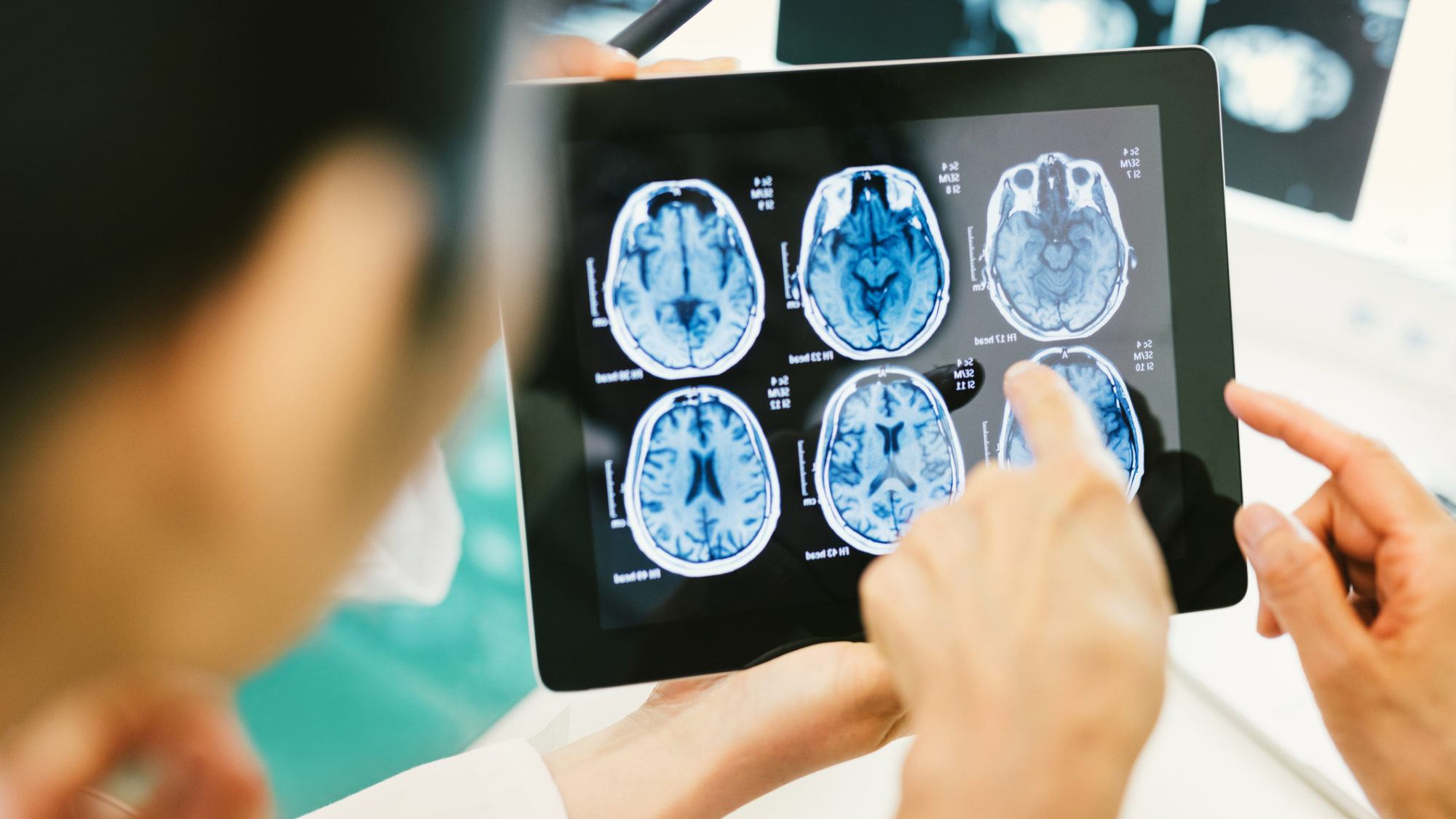
MEG detects the tiny magnetic fields generated by the electrical currents flowing through the brain’s neurons. These magnetic fields are measured using extremely sensitive devices known as SQUIDs (superconducting quantum interference devices), which can detect changes in magnetic fields that are billions of times smaller than the Earth’s magnetic field. Your epilepsy specialist can use data collected by MEG to create detailed maps of brain activity, often combined with structural imaging techniques like MRI (magnetic resonance imaging) to provide more comprehensive insights into brain function.
Because MEG is non-invasive and does not involve radiation, it can be safely used in various clinical and research settings. Barrow physicians use MEG for pre-surgical mapping of brain function in people with epilepsy, brain tumors, or other conditions requiring neurosurgery. In research, MEG is used to study the neural mechanisms underlying cognition, perception, and other psychological processes.

What is MEG used for?
MEG is used for several important applications, leveraging its exceptional temporal (time) resolution and good spatial (location) accuracy in mapping brain activity.
Our neurologists primarily use MEG for epilepsy evaluation and treatment planning. MEG is highly valuable in locating the regions of the brain where epileptic seizures originate. By identifying these zones, neurologists can better plan for surgical interventions to remove or reduce the area causing seizures while minimizing risks to critical brain functions. This is especially crucial for people whose seizures are not well-controlled by medication.
Additional uses for MEG include:
- Pre-surgical Brain Mapping: For patients needing surgery for tumors, arteriovenous malformations, or other brain abnormalities, MEG helps map out essential functions such as speech, motor control, and sensory processing. This mapping ensures surgeons can navigate these critical areas to reduce the risk of post-surgical deficits.
- Functional Brain Disorders: MEG is used to study and understand various functional brain disorders, including autism, Alzheimer’s disease, schizophrenia, and others. It can help understand the neural mechanisms underlying these conditions and contribute to developing new therapeutic strategies.
- Cognitive Neuroscience Research: Although not strictly clinical, MEG is widely used to understand how the brain processes information, including perception, language, memory, and emotion.
- Assessment of Brain Injuries: MEG can be used to analyze brain function in people who have suffered head injuries or have conditions like stroke. By examining the brain’s magnetic activity, neurologists can understand the extent of the damage and the affected brain areas, which can guide rehabilitation efforts.
Overall, MEG’s ability to provide real-time, dynamic images of brain activity makes it a powerful tool in the diagnosis, treatment planning, and research of neurological conditions. Its non-invasive nature also makes it an attractive option for repeated assessments, allowing for monitoring disease progression or treatment effects.
Am I a good candidate for MEG?
You may be a good candidate for Magnetoencephalography (MEG) if you have been diagnosed with epilepsy but do not respond well to medication and are considering surgery. MEG helps localize the epileptogenic zone (the part of your brain that generates seizures) more precisely than other non-invasive methods, aiding in planning surgery to remove or lessen the area causing seizures while minimizing impact on critical brain functions.
Other candidates for MEG could include:
- People with Brain Tumors: Before surgery to remove a brain tumor, MEG can be used for functional mapping to identify areas responsible for essential functions like speech, movement, and sensory processing. This helps surgeons avoid these critical areas during tumor removal.
- Individuals with Brain Lesions due to Stroke or Trauma: MEG can be employed to assess the functional impact of lesions and guide rehabilitation strategies by providing insights into how brain activity has been altered or reorganized following injury.
- Candidates for Neurosurgical Procedures: Beyond tumor resection and epilepsy surgery, MEG can assist in planning surgeries involving areas of the brain responsible for critical functions, preserving these areas.
- Research Participants: Individuals participating in cognitive neuroscience research, especially studies on how the brain processes information (like language, memory, and perception), can be good candidates for MEG. This research often seeks to understand the timing and sequencing of brain activity, areas where MEG excels.
- People with Functional Brain Disorders: Although more exploratory, MEG can be used to study conditions such as autism spectrum disorder, Alzheimer’s disease, schizophrenia, and other neurological or psychiatric conditions. It may help in understanding the neurobiological underpinnings of these disorders.
You should discuss with your healthcare providers whether MEG is appropriate for your specific situation, considering the detailed information needed, the condition being treated or studied, and how MEG results might influence your treatment plan or contribute to research outcomes.
Common Questions
What is the difference between MEG and EEG?
Both Magnetoencephalography (MEG) and Electroencephalography (EEG) are techniques used to measure brain activity, but they do so in different ways.
EEG (Electroencephalography)
- Measures the electrical activity of the brain using electrodes placed on the scalp
- Detects the small electrical currents generated by the firing of neurons
- Provides information about the timing and patterns of brain activity
- More widely available and less expensive than MEG
- Limited spatial resolution (ability to pinpoint activity to specific brain regions)
MEG (Magnetoencephalography)
- Measures the magnetic fields produced by the electrical activity of the brain
- Detects the tiny magnetic fields using sensitive sensors placed around the head
- Offers excellent spatial resolution, allowing for precise localization of brain activity
- Less affected by the skull and scalp compared to EEG, providing clearer signals
- Generally less widely available than EEG (for example, the only MEG device in Arizona is located at Barrow)
In summary, while both EEG and MEG provide valuable information about brain activity, MEG offers higher spatial resolution and is often used when precise localization of brain activity is needed, such as in surgical planning or research studies requiring detailed mapping of brain function.
Information and Resources
References
- Foldes ST, Chandrasekaran S, Camerone J, Lowe J, Ramdeo R, Ebersole J, Bouton CE. Case Study: Mapping Evoked Fields in Primary Motor and Sensory Areas via Magnetoencephalography in Tetraplegia. Front Neurol. 2021 Sep 24;12:739693. doi: 10.3389/fneur.2021.739693. PMID: 34630308; PMCID: PMC8497881.
- Shvarts V, Mäkelä JP. Auditory Mapping With MEG: An Update on the Current State of Clinical Research and Practice With Considerations for Clinical Practice Guidelines. J Clin Neurophysiol. 2020 Nov;37(6):574-584. doi: 10.1097/WNP.0000000000000518. PMID: 33165230.
- Shvarts V, Zoltay G, Bowyer SM, Zillgitt A, Moran JE, Mason K, Tepley N, Burdette D. Periodic Discharges: Insight From Magnetoencephalography. J Clin Neurophysiol. 2017 May;34(3):196-206. doi: 10.1097/WNP.0000000000000356. PMID: 27832046.


